

Evaluating Heart Function Under Stress
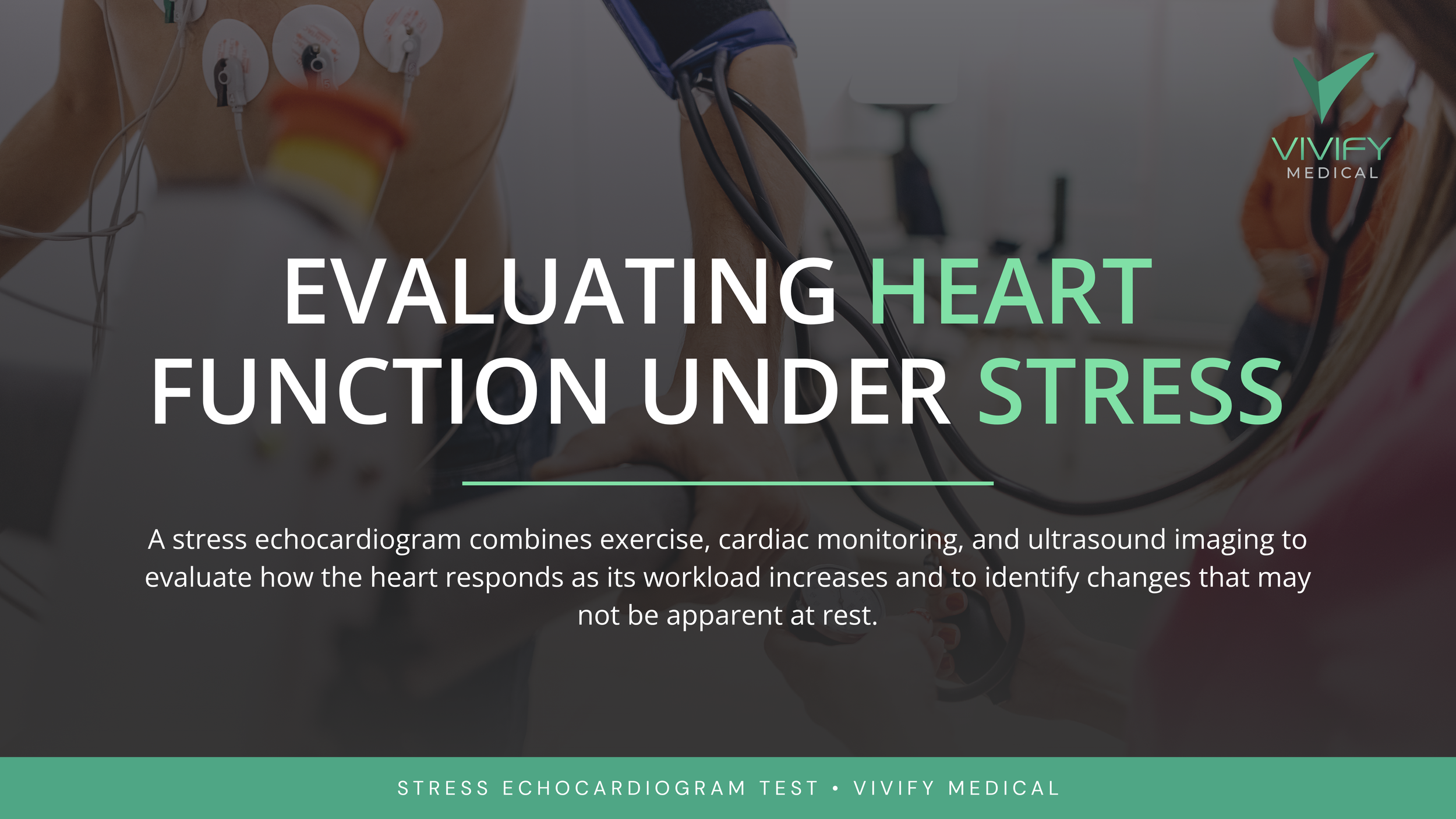
Some heart conditions are difficult to spot when your body is at rest. A stress echocardiogram combines physical exercise, ultrasound imaging, and electrocardiogram (ECG) tracking to evaluate how your heart performs as its workload rises. By comparing rest and exercise images, cardiologists can see how effectively your heart muscle contracts and check for signs of reduced blood flow.
1. Why evaluate the heart under stress?
Your heart needs a steady supply of oxygen, and that demand spikes during physical activity as it beats faster and pumps harder to supply your body. This relationship is called myocardial oxygen demand.
Healthy coronary arteries expand to deliver extra blood flow when demand rises. However, if an artery is narrowed, blood flow cannot keep up during exertion.
Because this mismatch often goes unnoticed while you are resting, an exercise stress test allows your cardiologist to see how your heart handles real-time physical demand. Adding ultrasound imaging allows us to directly compare your heart muscle's contractions at rest versus during exercise.
2. What a stress echocardiogram reveals
Comparing resting images with stress images highlights how different areas of your heart muscle perform under pressure:
Regional Wall Motion Abnormalities: If a specific region of the heart contracts less effectively during exercise, it may point to inducible ischemia—meaning that section of the heart muscle isn't receiving enough blood flow to meet its oxygen needs.
Electrical & Systemic Responses: The ECG monitors electrical activity for exercise-induced changes, while your care team tracks your vitals, physical comfort, and overall exercise capacity.
Symptom Mapping: Chest discomfort, abnormal blood pressure spikes, or sudden fatigue help identify underlying issues with physical conditioning or cardiovascular risk.
Your cardiologist interprets these results alongside your health history and risk factors to tailor a plan, whether that involves lifestyle modifications, medication updates, or targeted follow-up care.
3. What to expect during the test
A stress echocardiogram seamlessly pairs physical activity with real-time monitoring across four straightforward steps:
Resting Baseline: Small electrodes are placed on your chest to record your baseline ECG, heart rate, and blood pressure. We then perform a baseline ultrasound image of your resting heart.
Controlled Exercise: You will walk on a treadmill as the speed and incline gradually increase. Your team continuously monitors your heart rate, rhythm, blood pressure, and comfort level.
Stress Imaging: Right at peak exertion, we obtain a second set of ultrasound images to capture how your heart muscle contracts under maximum workload.
Monitored Recovery: You will rest comfortably while your vitals return to normal. (Note: The test can be modified or stopped at any time based on your symptoms or safety goals.)
4. Stress echocardiography at Vivify Medical
At Vivify Medical in Lower Manhattan, stress echocardiography forms an essential pillar of our preventive cardiovascular care. Combining advanced imaging with monitored exercise lets us uncover subtle heart issues that remaining still simply won't show.
Our cardiology team evaluates your test results within the full context of your lifestyle, medical history, and personal wellness goals. Whether you are addressing new symptoms during exercise, managing risk factors, or aiming to extend your healthspan by protecting the years you spend active and free from major disease, our team provides actionable steps forward.
Schedule a Consultation
If you are experiencing chest tightness, shortness of breath during workouts, or a change in exercise tolerance, contact Vivify Medical today to evaluate whether a stress echocardiogram is right for you.